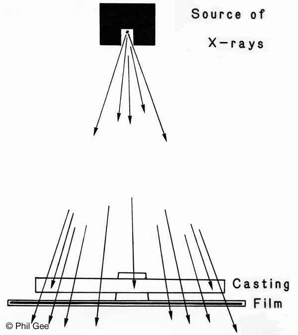
A Radiograph is an image created without the use of a conventional camera, lens or negative on photosensitive material, it employs wave lengths from the more energetic end of the electromagnetic spectrum: X-rays and Gamma radiation being the most common used today.
Industrial Radiography
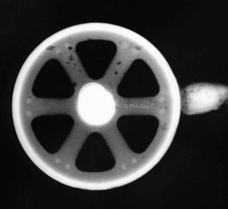
One can see from visual inspection of the plate defects in the spokes of the casting, shown by dark areas.
The Rim and center Boss which are thick show up white having absorbed all the radiation, whereas the spokes being thinner have absorbed less and show up gray. The defects probably gas pockets in the castings have absorbed less so show up as dark blobs. There may be defects in the thick areas but other techniques will have to be used to identify these.
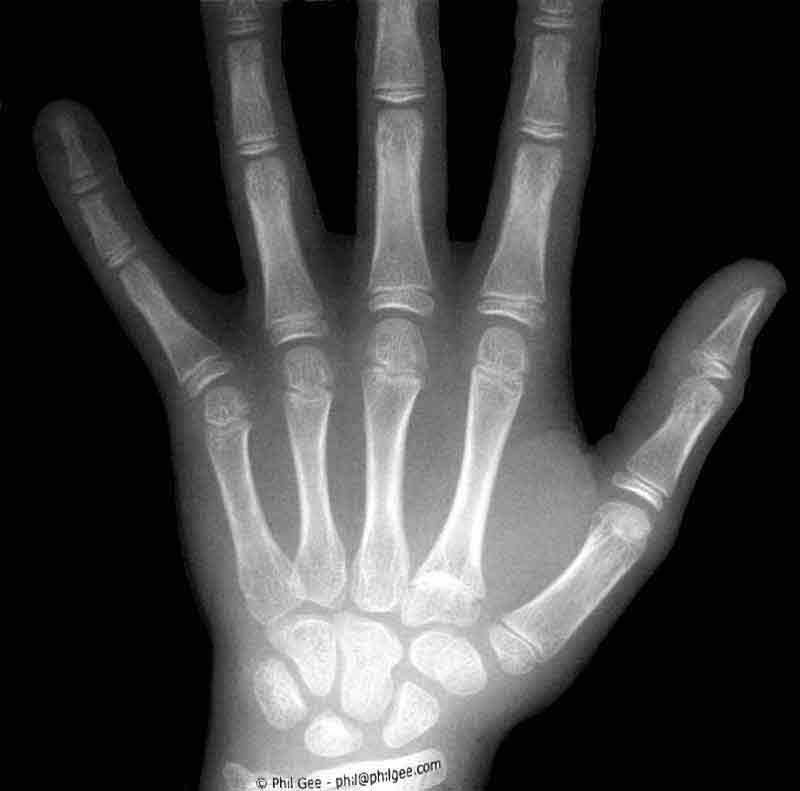
Medical Radiography
The resulting image is a 'negative' in the conventional sense where the dark areas represent maximum exposure the white areas minimum and the grays proportional amounts dependent upon the density, opacity, refractivity of the subject and the amount of exposure given.
However modern day X-ray techniques use digital imaging with the plate image being displayed on a digital monitor.
In a CT scan ( Computed tomography ) a three dimensional image is computed from a series of X-ray images of 'slices' through the object under investigation